

Plant root border cells are living, terminally differentiated cells produced by the root cap and released into the rhizosphere as single cells, sheets, or aggregates depending on species and developmental context. They aren’t just sloughed off, rather they are developmentally programmed, tied to targeted cell-wall remodeling and secretory capacity that persists after release. Functionally, this creates a mobile, renewable boundary layer that can be replenished when removed, allowing the plant to maintain an active biochemical interface between meristematic tissue at the root tip and the soil microbiome (Hawes et al., 1998). More recent synthesis provided evidence that the detachment of border cell, mucilage composition, and secretory outputs vary across taxa and environmental conditions, making border cells best understood as a regulated interface system rather than a single conserved phenotype (Omelyanchuk et al., 2024; Kranawetter & Sumner, 2025).
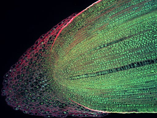
Maize root tip, by J. Haseloff, https://www.flickr.com/photos/71183136@N08/7093253363
Inducible investment in border cells
The work of Curlango-Rivera et al. (2010) on pea (Pisum sativum) showed that with exposure to pisatin, a phytoalexin, border-cell numbers increased, while exposure to ferulic acid, a common phenylpropanoid associated with cell-wall matrices, suppressed border-cell production. This suggests that the plant may choose to invest in a thicker biochemical boundary when defensive metabolites indicate an activated defense state, while damage-associated chemistry where cell wall fragments are present shifts priorities away from border-cell proliferation. This type of tradeoff is compatible with the general view of border cells as a controllable interface system rather than a fixed anatomical byproduct (Hawes et al., 1998; Kranawetter & Sumner, 2025).
Using integrated metabolomics and transcriptomics analysis of Medicago truncatula Watson et al. (2015) describes a linked pattern of broad downshifts in primary metabolism transcripts alongside large increases in specialized-metabolic pathways, including flavonoids and triterpenoid saponins. This suggests that border cells are pre-armed with defensive and signaling chemistry to protect the meristem and shape microbial interactions.
Border-cell exudation as chemical-ecology
Border cells secrete complex mixtures of carbohydrates, proteins, amino acids, organic acids, extracellular DNA, and specialized metabolites into mucilage, thereby shaping microbial recognition, attachment, growth, and spatial organization near the root tip. Mechanistically, this matters because the ecological problem is not just the existence of pathogens, but that the rhizosphere is a carbon-limited arena where microbes compete for plant-derived substrates and for access to colonization sites. Border-cell mucilage and exudates alter that competitive landscape by changing resource availability, adhesion, and antimicrobial pressure in a highly localized zone. Reviews spanning multiple systems converge on the idea that border cells can simultaneously attract beneficial microbes and constrain pathogens through selective chemistry embedded in mucilage matrices (Hawes et al., 1998; Omelyanchuk et al., 2024; Fortier et al., 2023).
Shirakawa et al., (2023) described the concept of a root extracellular trap to reframe border cells plus mucilage as an organized defensive structure that combines physical entanglement with chemical and nucleic-acid mediated effects. Their work in woody systems adds a modern, visually grounded in Pinus densiflora roots of an extracellular trap that interacts with root-associated bacteria. They provide evidence that trapped bacteria can induce reactive oxygen species responses in cap-derived cells. This supports the idea that the trap is not simply a sterilizing barrier but a selective filter that can foster a protective consortium under some conditions.
Border-cell signaling as chemical-ecology
Microbes can actively modulate the chemical environment they inhabit, potentially shifting the composition of the root-interface metabolite pool that includes border-cell exudate (Phillips et al., 2004). Specific secondary metabolites, especially flavonoids and isoflavonoids, serve as both antimicrobial/defensive compounds and symbiotic signals, shaping fungal behavior and plant transcriptional programs during colonization. Primary evidence in Medicago truncatula shows that mycorrhizal establishment is associated with shifts in isoflavonoid accumulation and defense-gene transcripts, consistent with a signaling role for phenylpropanoid-derived metabolites in symbiosis (Harrison & Dixon, 1993). However, the proportion of total symbiotic signaling that is specifically border-cell derived requiring spatially resolved sampling (Kranawetter & Sumner, 2025; Harrison & Dixon, 1993).
A 2024 study comparing five medicinal plants found that root exudate metabolite profiles differ by species and correlate with distinct bacterial and fungal community profiles, identifying specific metabolites associated with microbial shifts (Qu et al., 2024). A 2025 study in Rheum tanguticum explicitly links developmental-stage shifts in root exudates to rhizosphere microbiome changes and to measured differences in root bioactive compounds that suggest a cascade effect (Li et al., 2025). These studies do not isolate border cells as the sole source of exudates, but they strengthen the broader premise that root-interface chemistry can be a determinant of medicinal-plant phytochemical quality.
Systemic effects and root-to-shoot chemistry
Root apical regions show robust local communication, yet long-distance signaling pathways that connect root-zone events to whole-plant chemistry are not always straightforward. An example of systemic coupling is found in root-associated microbes reshaping the leaf metabolome and altering insect feeding behavior, which demonstrates that root microbiome context can propagate to aboveground chemistry (Badri et al., 2013).
Separately, work in Arabidopsis supports a bidirectional framework where the root microbiome can modulate root exudation, implying feedback loops between microbial community structure and plant chemical outputs at the root interface (Korenblum et al., 2020). Together, these studies support the idea that the rhizosphere can influence distal phytochemical phenotypes, but the transport forms and signaling intermediates that connect border-cell chemistry to leaf metabolomes remain incompletely specified and should not be assumed to be direct border-cell to leaf transport without targeted tracing (Badri et al., 2013; Korenblum et al., 2020; Turnbull & Lopez-Cobello, 2013).
References
Badri, D. V., Zolla, G., Bakker, M. G., Manter, D. K., & Vivanco, J. M. (2013). Potential impact of soil microbiomes on the leaf metabolome and on herbivore feeding behavior. New Phytologist, 198(1), 264–273. https://doi.org/10.1111/nph.12124
Curlango-Rivera, G., Duclos, D. V., Clay, N. K., & Hawes, M. C. (2010). Root border cells protect root tips from pathogens and toxic metals. Plant and Soil, 336, 5–17. https://doi.org/10.1007/s11104-010-0291-8
Fortier, M., Lemaitre, V., Gaudry, A., Pawlak, B., Driouich, A., & Follet-Gueye, M.-L. (2023). A fine-tuned defense at the pea root caps: Involvement of border cells and arabinogalactan proteins against soilborne diseases. Frontiers in Plant Science, 14, 1132132. https://doi.org/10.3389/fpls.2023.1132132
Harrison, M. J., & Dixon, R. A. (1993). Isoflavonoid accumulation and expression of defense gene transcripts during the establishment of vesicular-arbuscular mycorrhizal associations in roots of Medicago truncatula. Molecular Plant-Microbe Interactions, 6, 643–654. https://doi.org/10.1094/MPMI-6-643
Hawes, M. C., Brigham, L. A., Wen, F., Woo, H. H., & Zhu, Y. (1998). Function of root border cells in plant health: Pioneers in the rhizosphere. Annual Review of Phytopathology, 36, 311–327. https://doi.org/10.1146/annurev.phyto.36.1.311
Hawes, M. C., Bengough, G., Cassab, G., & Ponce, G. (2002). Root caps and rhizosphere. Journal of Plant Growth Regulation, 21, 352–367. https://doi.org/10.1007/s00344-002-0035-y
Kape, R., Wex, K., Parniske, M., Görge, E., Wetzel, A., & Werner, D. (1993). Legume root metabolites and VA-mycorrhiza development. Journal of Plant Physiology, 141(1), 54–60. https://doi.org/10.1016/S0176-1617(11)80851-5
Korenblum, E., Dong, Y., Szymanski, J., Panda, S., Jozwiak, A., Massalha, H., Meir, S., Rogachev, I., Aharoni, A. (2020). Rhizosphere microbiome mediates systemic root metabolite exudation by root-to-root signaling. Proceedings of the National Academy of Sciences of the United States of America, 117(7), 3874–3883. https://doi.org/10.1073/pnas.1912130117
Kranawetter, C., & Sumner, L. W. (2025). Differential root zone secretions and the role of root border cells in rhizosphere manipulation. Phytochemistry Reviews, 24(6), 5639–5658. https://doi.org/10.1007/s11101-025-10084-y
Li, J., Wang, B., Ma, H., Yang, X., Wang, T., & Zhou, G. (2025). Root exudates drive plant-microbiome interactions influencing the quality of cultivated Rheum tanguticum during different growth development stages. Industrial Crops and Products, 237, 122157. https://doi.org/10.1016/j.indcrop.2025.122157
Omelyanchuk, N. A., Cherenko, V. A., & Zemlyanskaya, E. V. (2024). Root cap border cells as regulators of rhizosphere microbiota. Vavilovskii Zhurnal Genetiki i Selektsii, 28(8), 918–926. https://doi.org/10.18699/vjgb-24-99
Phillips, D. A., Fox, T. C., King, M. D., Bhuvaneswari, T. V., & Teuber, L. R. (2004). Microbial products trigger amino acid exudation from plant roots. Plant Physiology, 136(1), 2887–2894. https://doi.org/10.1104/pp.104.044222
Qu, P., Wang, B., Qi, M., Lin, R., Han, C., Xie, C., Zhang, Z., & Qiu, J. (2024). Medicinal plant root exudate metabolites shape the rhizosphere microbiota. International Journal of Molecular Sciences, 25(14), 7786. https://doi.org/10.3390/ijms25147786
Rüger, A., Girke, C., Lah, M., Borgmann, S., Banasiak, R., Drauschke, T., Strehmel, N., Scheel, D., & Herschbach, C. (2023). Bacterial root colonization reorganizes the Arabidopsis thaliana root transcriptome and exudation profile. New Phytologist, 238(3), 1372–1390. https://doi.org/10.1111/nph.19002
Shirakawa, M., Matsushita, N., & Fukuda, K. (2023). Visualization of root extracellular traps in an ectomycorrhizal woody plant (Pinus densiflora) and their interactions with root-associated bacteria. Planta, 258(6), 112. https://doi.org/10.1007/s00425-023-04274-1
Turnbull, L. A., & Lopez-Cobello, R. M. (2013). Communication and complexity in the root apical meristem. New Phytologist, 199(4), 871–873. https://doi.org/10.1111/nph.12167
Watson, B. S., Bedair, M. F., Urbanczyk-Wochniak, E., Huhman, D. V., Yang, D. S., Allen, S. N., Li, W., Tang, Y., & Sumner, L. W. (2015). Integrated metabolomics and transcriptomics reveal enhanced specialized metabolism in Medicago truncatula root border cells. Plant Physiology, 167(4), 1699–1716. https://doi.org/10.1104/pp.114.253054