

This and following posts will be serialized from a longer piece of writing. The focus will be broken up into sections:
Endophytic Transition States
Secondary Metabolites as System Equilibrium
Carbon Allocation Governs Chemical Expression
Signal Convergence
Wild Ecosystems
Ratio Control as Emergent
Finding Terroir
The prior sections describe a system in which regulation of secondary metabolism occurs across the plant–soil boundary. Signals originate in root exudates, microbial communities assemble in response, and feedback alters plant metabolic state. This configuration depends on distance. Signals must diffuse, microbial partners must localize to the rhizoplane, and responses are mediated across cellular interfaces. Endophytic colonization alters the relationship.
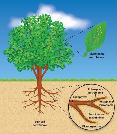

When fungi form internal, often asymptomatic associations within plant tissues they are referred to as endophytes. The fungus colonizes intercellular spaces or specific cell types while the host maintains normal growth. In many systems, these relationships influence plant metabolism and stress responses without causing disease. These endophytic fungi can modulate plant biochemical pathways from within plant tissues rather than through external rhizosphere signaling (Schardl et al., 2004; Clay & Schardl, 2002).
Endophytic colonization establishes direct biochemical and signaling interactions within plant tissues, reducing spatial separation between signal origin and response and enabling more immediate modulation of host processes (Hardoim et al., 2015; Brader et al., 2017). Endophytic colonization reduces spatial separation between microbial signals and host responses, enabling direct biochemical interactions within plant tissues (Hardoim et al., 2015; White et al., 2019).
Secondary metabolism, in this context, is influenced not only by external ecological structure but also by embedded biological components. Experimental studies show that endophytes can modulate host biosynthetic gene expression and associated metabolite accumulation, indicating that internal microbial presence contributes directly to regulation of secondary metabolic pathways rather than acting solely through external environmental cues (Kusari et al., 2012; Brader et al., 2017). This supports a model in which metabolic outcomes reflect integrated regulation across both external microbial communities and internalized symbiotic partners.
The transition from rhizosphere association to endophytic colonization can be viewed as a continuum rather than a discrete shift, as many microbes exhibit both lifestyles depending on environmental conditions and host state (Hardoim et al., 2015; Compant et al., 2010). Initial contact occurs at the root surface and exudate components influence microbial behavior and colonization (Hassan & Mathesius, 2012; Badri & Vivanco, 2009). Successful colonization involves modulation of plant immune responses, allowing entry into root tissues and establishment within intercellular or intracellular spaces (Hardoim et al., 2015).
Endophyte studies support bidirectional interaction between host metabolism and internal microbial activity, although direct demonstration that endophyte responses feedback to alter host metabolic state in this specific system is not yet available (White et al., 2019; Brader et al., 2017). In this context, metabolite production contributes to structuring endophytic community composition and activity, supporting a potential feedback relationship in which host biosynthetic output shapes internal microbial behavior that may, in turn, influence plant metabolic state.
This internalization has consequences for carbon allocation and nutrient exchange. Endophytes draw on host-derived carbon while contributing to nutrient acquisition, stress tolerance, or defense, depending on the system (Compant et al., 2010; Rodriguez et al., 2009). Because the interaction occurs within plant tissues, exchange processes are less influenced by bulk soil conditions and diffusion constraints, enabling more direct and potentially more regulated interactions between partners. This can increase interaction efficiency and stabilize effects on plant physiology and metabolism. In contrast, rhizosphere interactions remain subject to environmental variability, including fluctuations in soil moisture, temperature, and competing microbial populations, which can alter both microbial composition and functional outcomes (Bardgett & van der Putten, 2014).
The presence of endophytes also alters the plant’s perception of its environment. Internal microbes can prime defense pathways, leading to modified responses to subsequent stimuli, a process described as induced systemic resistance (ISR) (Pieterse et al., 2014). These priming influences secondary metabolism by maintaining components of defense-related pathways in a responsive state. Endophyte-associated signaling, similar to mycorrhizal association, overlaps with pathways that regulate secondary metabolite production, including jasmonate and salicylate signaling networks (Pieterse et al., 2012; Rodriguez et al., 2009). As a result, the baseline metabolic state of the plant can shift in the absence of immediate external stress,.
In Salvia miltiorrhiza, inoculation with the endophytic fungus Cladosporium tenuissimum enhanced tanshinone accumulation alongside upregulation of genes in the biosynthetic pathway, indicating coordinated transcriptional and metabolic responses to internal microbial presence (Chen et al., 2022c). Endophytic bacteria and fungi have been reported to enhance production of flavonoids, alkaloids, and terpenoids across plant systems as diverse as Taxus spp., Ginkgo biloba, and Hypericum perforatum (Pang et al., 2021).
Evidence specific to Echinacea purpurea indicates that this species hosts diverse endophytic bacterial communities, including Pseudomonas, Bacillus, and actinomycetes. Broader endophyte reviews support signaling-based effects of internal microbes on host physiology and metabolism, but direct demonstration in E. purpurea remains limited (Ryan et al., 2008; White et al., 2019). Experimental analyses of E. purpurea preparations further show that immunostimulatory activity is partly attributable to bacterial lipopolysaccharides and related microbe-associated molecular patterns present in extracts, indicating that observed bioactivity reflects contributions from both plant-derived compounds, such as alkamides and caffeic acid derivatives, and associated microbial components (Pugh et al., 2008).
Several compounds originally attributed to medicinal plants have been shown to be produced, or co-produced, by endophytic fungi associated with those plants. The most widely cited example is paclitaxel (taxol), first isolated from Taxus species and later reported from endophytic fungi such as Taxomyces andreanae (Stierle et al., 1993). Similar observations have been reported for other bioactive classes, including camptothecin from endophytes of Camptotheca acuminata and related hosts, and podophyllotoxin from endophytes associated with Podophyllum species (Kusari et al., 2012; Puri et al., 2005). In these systems, endophytes can contribute directly to the pool of bioactive metabolites or influence host metabolism, complicating attribution of compound origin. While not universal and sometimes difficult to reproduce across isolates, these findings demonstrate that endophytic fungi can be a source of pharmacologically relevant compounds traditionally assigned to plant biosynthesis (Kusari et al., 2012).
References
Badri, D. V., & Vivanco, J. M. (2009). Regulation and function of root exudates. Plant, Cell & Environment, 32(6), 666–681. https://doi.org/10.1111/j.1365-3040.2009.01926.x
Brader, G., Compant, S., Vescio, K., Mitter, B., Trognitz, F., Ma, L.-J., & Sessitsch, A. (2017). Ecology and genomic insights into plant-pathogenic and plant-nonpathogenic endophytes. Annual Review of Phytopathology, 55, 61–83. https://doi.org/10.1146/annurev-phyto-080516-035641
Chen, H., Chen, J., Qi, Y., Chu, S., Ma, Y., Xu, L., Lv, S., Zhang, H., Yang, D., Zhu, Y., Mans, D. R. A., & Liang, Z. (2022). Endophytic fungus Cladosporium tenuissimum DF11, an efficient inducer of tanshinone biosynthesis in Salvia miltiorrhiza roots. Phytochemistry, 194, 113021. https://doi.org/10.1016/j.phytochem.2021.113021
Clay, K., & Schardl, C. (2002). Evolutionary origins and ecological consequences of endophyte symbiosis with grasses. The American Naturalist, 160(S4), S99–S127. https://doi.org/10.1086/342161
Compant, S., Clément, C., & Sessitsch, A. (2010). Plant growth-promoting bacteria in the rhizo- and endosphere of plants: Their role, colonization, mechanisms involved and prospects for utilization. Soil Biology and Biochemistry, 42(5), 669–678. https://doi.org/10.1016/j.soilbio.2009.11.024
Hardoim, P. R., van Overbeek, L. S., & van Elsas, J. D. (2015). Properties of bacterial endophytes and their proposed role in plant growth. Trends in Microbiology, 23(8), 463–471. https://doi.org/10.1016/j.tim.2015.04.008
Hassan, S., & Mathesius, U. (2012). The role of flavonoids in root-rhizosphere signalling: Opportunities and challenges for improving plant-microbe interactions. Journal of Experimental Botany, 63(9), 3429–3444. https://doi.org/10.1093/jxb/err430
Kusari, S., Hertweck, C., & Spiteller, M. (2012). Chemical ecology of endophytic fungi: Origins of secondary metabolites. Chemistry & Biology, 19(7), 792–798. https://doi.org/10.1016/j.chembiol.2012.06.004
Li, J., Zhao, G.-Z., Varma, A., Qin, S., Xiong, Z., Huang, H.-Y., Zhu, W.-Y., Zhao, L.-X., Xu, L.-H., Zhang, S., & Li, W.-J. (2012). An endophytic Pseudonocardia species induces the production of artemisinin in Artemisia annua. PLoS ONE, 7(12), e51410. https://doi.org/10.1371/journal.pone.0051410
Pang, Z., Chen, J., Wang, T., Gao, C., Li, Z., Guo, L., Xu, J., & Cheng, Y. (2021). Linking plant secondary metabolites and plant microbiomes: A review. Frontiers in Plant Science, 12, 621276. https://doi.org/10.3389/fpls.2021.621276
Pieterse, C. M. J., Van der Does, D., Zamioudis, C., Leon-Reyes, A., & Van Wees, S. C. M. (2012). Hormonal modulation of plant immunity. Annual Review of Cell and Developmental Biology, 28, 489–521. https://doi.org/10.1146/annurev-cellbio-092910-154055
Pieterse, C. M. J., Zamioudis, C., Berendsen, R. L., Weller, D. M., Van Wees, S. C. M., & Bakker, P. A. H. M. (2014). Induced systemic resistance by beneficial microbes. Annual Review of Phytopathology, 52, 347–375. https://doi.org/10.1146/annurev-phyto-082712-102340
Pugh, N. D., Tamta, H., Balachandran, P., Wu, X., Howell, J., Dayan, F. E., & Pasco, D. S. (2008). The majority of in vitro macrophage activation exhibited by Echinacea preparations is due to bacterial lipoproteins and lipopolysaccharides. Journal of Agricultural and Food Chemistry, 56(22), 10552–10556. https://doi.org/10.1021/jf8019158
Puri, S. C., Nazir, A., Chawla, R., Arora, R., Riyaz-ul-Hasan, S., Amna, T., Ahmed, B., Verma, V., Singh, S., Sagar, R., & Sharma, A. (2005). The endophytic fungus Entrophospora infrequens produces camptothecin. Journal of Natural Products, 68(12), 1717–1719. https://doi.org/10.1021/np0502802
Rodriguez, R. J., White, J. F., Arnold, A. E., & Redman, R. S. (2009). Fungal endophytes: Diversity and functional roles. New Phytologist, 182(2), 314–330. https://doi.org/10.1111/j.1469-8137.2009.02773.x
Ryan, R. P., Germaine, K., Franks, A., Ryan, D. J., & Dowling, D. N. (2008). Bacterial endophytes: Recent developments and applications. FEMS Microbiology Letters, 278(1), 1–9. https://doi.org/10.1111/j.1574-6968.2007.00918.x
Schardl, C. L., Leuchtmann, A., & Spiering, M. J. (2004). Symbioses of grasses with seedborne fungal endophytes. Annual Review of Plant Biology, 55, 315–340. https://doi.org/10.1146/annurev.arplant.55.031903.141735
Stierle, A., Strobel, G., & Stierle, D. (1993). Taxol and taxane production by Taxomyces andreanae, an endophytic fungus of Pacific yew. Science, 260(5105), 214–216. https://doi.org/10.1126/science.8097061
White, J. F., Kingsley, K. L., Zhang, Q., Verma, R., Obi, N., Dvinskikh, S., Elmore, M. T., Verma, S. K., & Kowalski, K. P. (2019). Review: Endophytic microbes and their potential applications in crop management. Plant and Soil, 438, 1–17. https://doi.org/10.1007/s11104-018-3811-3