

This will be a serial conversation about the following:
How is it collected and what are the active phytochemicals?
How is it extracted, sustainably sourced and processed?
Balancing extraction, stability, and skin delivery
Wounding (Tapping)
Within the living plant, resin exposure following wounding initiates a rapid physical and biochemical response. Volatile monoterpenes such as α-pinene and limonene are lost through evaporation on a timescale of hours to days, as demonstrated in conifer resin systems where monoterpene emissions peak immediately after injury and decline sharply within 24–72 hours (Loreto et al., 2000; Niinemets et al., 2013). Volatile monoterpenes appear to be partially retained within the viscous resin matrix, where increasing viscosity, hydrophobic association, and progressive oxidative cross-linking slow diffusion and volatilization over time (Langenheim, 2003; Turek & Stintzing, 2013).
Wounding is the primary trigger for resin exudation. Mechanical injury releases pre-formed resin immediately but also activates induced defense pathways mediated by jasmonic acid signaling, increasing terpenoid biosynthesis over time (Howe & Jander, 2008). This process induces terpene biosynthesis, with transcriptional upregulation of terpene synthase genes occurring within hours and persisting for several days, leading to altered terpene composition in newly produced resin (Martin et al., 2002; Keeling & Bohlmann, 2006).
Herbivory produces a distinct response relative to mechanical wounding because insect-derived elicitors activate additional signaling pathways, leading to altered terpene and secondary metabolite profiles beyond those induced by physical injury alone (Howe & Jander, 2008; Mithöfer & Boland, 2012). Mechanical wounding typically elevates constitutive monoterpenes and simple green-leaf volatiles released from damaged tissue, whereas herbivory often induces additional oxidized terpenoids, sesquiterpenes, and signaling volatiles such as linalool, β-caryophyllene, methyl salicylate, DMNT, and TMTT through insect-specific defense pathways.
For harvesting, controlled mechanical wounding is more reproducible than herbivore damage and avoids introducing additional biotic stress responses that may alter resin chemistry.
After harvest, enzymatic activity rapidly diminishes, and compositional changes are dominated by abiotic processes. Monoterpene loss continues through volatilization, with essential oil studies showing substantial reduction in monoterpene content within days to weeks depending on storage conditions, particularly under exposure to air and elevated temperature (Turek & Stintzing, 2013). Nonvolatile resin components undergo oxidative modification and progressive formation of higher–molecular weight, cross-linked triterpenoid aggregates, resulting in a less soluble and more rigid resin matrix. Studies of resin aging and terpenoid oxidation indicate that these processes proceed over weeks to months, increasing molecular weight distribution and viscosity while reducing solubility and extractability (Hemingway & McGraw, 1983; Langenheim, 2003).
Over longer storage periods, continued oxidation and cross-linking reactions can produce substantial physical changes. Fossilization and maturation studies of plant resins show that progressive polymerization and oxidation over months to years lead to hardening, embrittlement, and loss of volatile fractions, although the exact rate depends strongly on oxygen exposure, light, and temperature (Bray & Anderson, 2009). These processes are physicochemical rather than biological and result in material that differs significantly in extraction behavior from freshly exuded resin. The precise chemical pathways in Boswellia resin remain less resolved than the broader aging behavior of plant resins.
Active Compounds and Structure-Function Relevance
The primary compounds of interest in Boswellia are boswellic acids, particularly 11-keto-β-boswellic acid and 3-acetyl-11-keto-β-boswellic acid (Fig. 3), along with related ursane- and oleanane-type triterpenoids (Rehman et al., 2025).
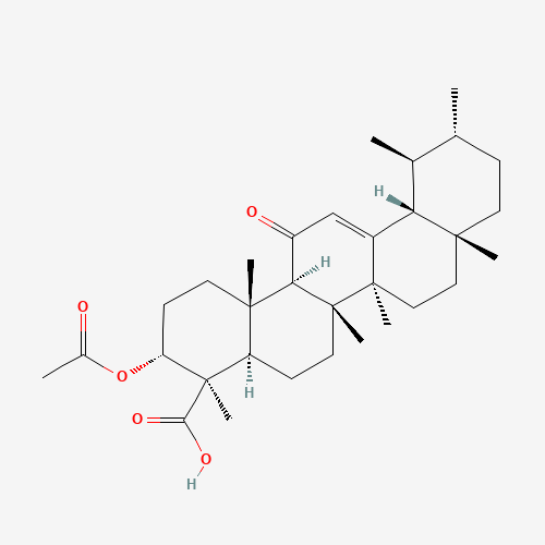
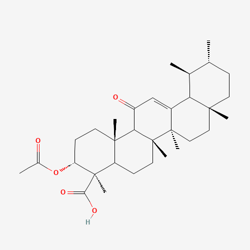
Acetyl-11-Keto-Beta-Boswellic Acid
3-Acetyl-11-Keto-Beta-Boswellic Acid
Figure 3. Boswellic acid structural features relevant to solubility and biological activity.
The C-11 keto group, C-3 acetylation, and carboxyl group affect polarity, membrane partitioning, and interaction with 5-lipoxygenase.
Boswellic acids are formed through cyclization of squalene followed by oxidative modification, producing the characteristic pentacyclic triterpenoid structures (Rehman et al., 2025) with minimal polar surface area, limited ionization at physiological pH (Roy et al., 2019). These molecules are large, rigid, and highly lipophilic, which limits their solubility in aqueous environments and complicates both extraction and delivery (Siddiqui, 2011).
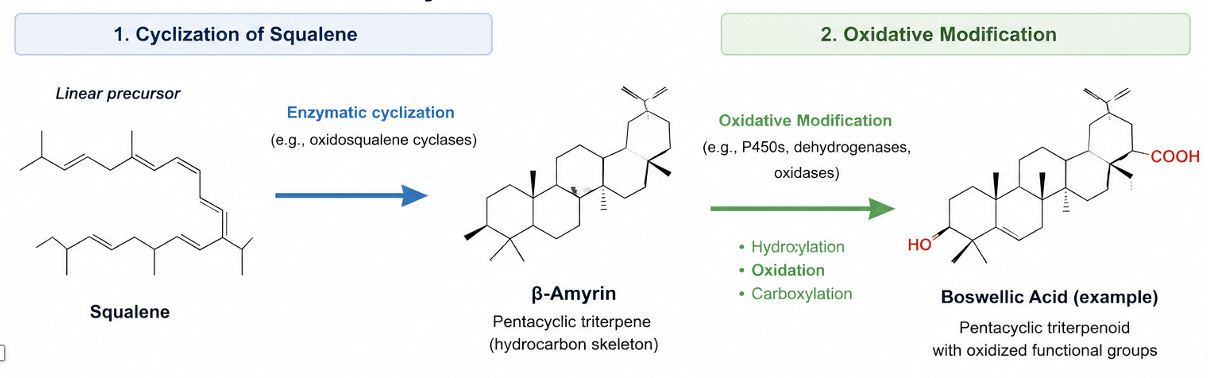
Figure 4: Formation of boswellic acids from squalene through cyclization and oxidation.
The stratum corneum (outer most layer of skin) is a lipid-rich barrier composed primarily of ceramides, cholesterol, and fatty acids. Because the stratum corneum is lipid-rich, lipophilic compounds such as boswellic acids are expected to partition into outer skin layers, but movement into deeper viable tissue is limited by molecular size and low aqueous solubility (Nilius & Bíró, 2013; Steinhoff et al., 2014). However, further movement into the viable epidermis and dermis is restricted by both molecular size and low aqueous solubility, limiting passive diffusion beyond the superficial barrier (Roy et al., 2019). As a result, penetration into deeper tissue layers depends on the ability of the formulation to facilitate transfer across both lipid and aqueous domains (Mostafa et al., 2015; Priya et al., 2025).
Systemic pharmacokinetic studies show low plasma concentrations and strong lipid association following oral dosing of boswellic acids, reinforcing the constraint that distribution is governed by lipid partitioning rather than free diffusion (Hüsch et al., 2013; Skarke et al., 2012). In a Boswellia carterii study of in vitro and in vivo transdermal permeation study, an applied microemulsion system with droplet sizes below 100 nm and encapsulation efficiencies above 83%, found that the lower-oil microemulsion displayed higher permeation parameters than the higher-oil comparator (Mostafa et al., 2015).
A second formulation optimization and ex vivo permeation study used 3-acetyl-11-keto-β-boswellic acid (AKBA). The results demonstrated that an AKBA-loaded nanostructured lipid carrier gel increased ex vivo skin permeation 30% more than AKBA gel alone while maintaining measurable skin retention. In both studies, boswellic-acid skin delivery improved when the delivery vehicle was engineered to overcome the hydrophobicity-driven release barrier (Priya et al., 2025).
At the target level, the rigid, hydrophobic structure of boswellic acids is consistent with interaction at lipid–protein interfaces, including membrane-associated enzymes involved in inflammatory signaling. In vitro work demonstrates inhibition of 5-lipoxygenase and related pathways (Roy et al., 2019). The C-11 keto group, C-3 acetylation, and carboxyl group alter polarity, hydrogen-bonding capacity, and membrane partitioning, which helps explain why boswellic acid analogues differ in both bioavailability and 5-lipoxygenase inhibitory activity (Sailer et al., 1996).
These features also affect interaction with lipid carriers, where increased lipophilicity enhances incorporation into oil phases but may reduce release from the formulation, so vehicle design becomes a major determinant of actual skin delivery (Mostafa et al., 2015; Priya et al., 2025). This creates a formulation limit where extraction efficiency and delivery efficiency are not aligned. Maximizing extraction into oil does not necessarily maximize bioavailability at the site of action, because retention in the carrier and transfer into skin are separate steps.
Practical strategies to increase release without losing the carrier’s absorption benefit include using mixed lipid carriers rather than a single oil phase, adding phospholipids or surfactants to improve partitioning, reducing particle size to increase contact with the stratum corneum, using deformable vesicles or nanostructured lipid carriers to improve skin contact, and tuning the oil-to-surfactant ratio so the compound is not trapped too tightly in the vehicle (Hüsch et al., 2012; Mehta et al., 2016; Priya et al., 2025; Sharma et al., 2010; Varia et al., 2022).
Potentially active compounds found in the extract are not limited to boswellic acids. Other triterpenoids and minor constituents contribute to overall activity. Isolated from Boswellia sacra (Fig. 5), cembranoids have been shown to act as TRPV3 agonists, suggesting a sensory or neuromodulatory component that may contribute to topical effects (Al-Harrasi et al., 2021). Boswellia extracts may alter how discomfort is perceived at the skin level, independent of boswellic acid–driven anti-inflammatory effects (Zhang et al., 2026).
Cembranoids are defined as macrocyclic diterpenoids (C20 isoprenoids) possessing a 14-membered cembrane ring. Many reported derivatives are oxygenated (e.g., hydroxylated, epoxidized, or lactonized) with changes to associated biological activity including receptor modulation and anti-inflammatory effects (Li et al., 2013; Wahlberg & Eklund, 1992).
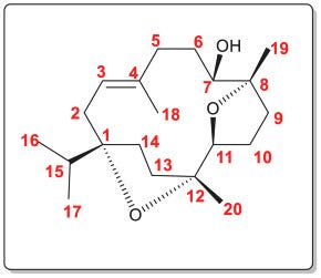
Figure 5. Oxygenated cembranoids from Boswellia sacra and possible TRPV3 activity.
Minor diterpenoids may contribute to skin-level sensory effects distinct from boswellic-acid-driven anti-inflammatory mechanisms [Al-Harrasi et al. (2021) https://doi.org/10.1016/j.phytochem.2021.112897]
Because transient receptor potential vanilloid 3 (TRPV3) channels are expressed primarily in keratinocytes within superficial skin layers, compounds do not need to penetrate deeply to produce functional results (Nilius & Bíró, 2013; Steinhoff et al., 2014). TRPV3 activation may contribute to sensory effects in skin, but the direction of that effect depends on dose, exposure duration, and compound structure. Strong or prolonged activation may contribute to irritation rather than relief. TRPV3 signaling in skin cells can also influence local mediator release, linking sensation with changes in the local inflammatory environment (Nilius & Bíró, 2013; Steinhoff et al., 2014). This supports the interpretation that some components of Boswellia extracts may contribute to symptomatic relief through localized sensory modulation at the skin surface.
In the Boswellia sacra study, TRPV3 activity was associated with structurally modified cembranoids rather than the bulk nonpolar fraction, supporting the possibility that minor oxygenated diterpenoids contribute to skin-level sensory effects (Nilius & Bíró, 2013; Steinhoff et al., 2014; Zhang et al., 2026). This supports the interpretation that the sensory or neuromodulatory effects attributed to Boswellia extracts are likely driven by a subset of structurally modified diterpenoids rather than the bulk nonpolar fraction.
References
Al-Harrasi, A., Avula, S. K., Csuk, R., & Das, B. (2021). Cembranoids from Boswellia species. Phytochemistry, 191, 112897. https://doi.org/10.1016/j.phytochem.2021.112897
Bray, P. S., & Anderson, K. B. (2009). Identification of carbon–carbon double bond position in resin diterpenoids using pyrolysis-gas chromatography/mass spectrometry. Journal of Analytical and Applied Pyrolysis, 85(1–2), 54–59. https://doi.org/10.1016/j.jaap.2008.11.006
DeCarlo, A., Johnson, S., Poudel, A., Satyal, P., Bangerter, L., & Setzer, W. N. (2018). Chemical variation in essential oils from the oleo-gum resin of Boswellia carteri: A preliminary investigation. Chemistry & Biodiversity, 15(6), e1800047. https://doi.org/10.1002/cbdv.201800047
Hemingway, R. W., & McGraw, G. W. (1983). Kinetics of acid-catalyzed reactions of flavonoid model compounds. Journal of Wood Chemistry and Technology, 3(3), 421–431. https://doi.org/10.1080/02773818308085112
Howe, G. A., & Jander, G. (2008). Plant immunity to insect herbivores. Annual Review of Plant Biology, 59, 41–66. https://doi.org/10.1146/annurev.arplant.59.032607.092825
Hüsch, J., Bohnet, J., Fricker, G., Skarke, C., Artaria, C., Appendino, G., Schubert-Zsilavecz, M., & Abdel-Tawab, M. (2013). Enhanced absorption of boswellic acids by a lecithin delivery form (Phytosome®) of Boswellia extract. Planta Medica, 79(6), 431–435. https://doi.org/10.1055/s-0032-1328264
Hüsch, J., Gerbeth, K., Fricker, G., Setzer, C., Zirkel, J., Rebmann, H., Schubert-Zsilavecz, M., & Abdel-Tawab, M. (2012). Effect of phospholipid-based formulations of Boswellia serrata extract on the solubility, permeability, and absorption of the individual boswellic acid constituents present. Journal of Natural Products, 75(10), 1675–1682. https://doi.org/10.1021/np300009w
Keeling, C. I., & Bohlmann, J. (2006). Diterpene resin acids in conifers. Phytochemistry, 67(22), 2415–2423. https://doi.org/10.1016/j.phytochem.2006.08.011
Langenheim, J. H. (2003). Plant resins: Chemistry, evolution, ecology, and ethnobotany. Timber Press.
Li, Y., Pattenden, G., & Webb, R. (2013). Biomimetic and synthetic studies on cembranoid natural products. Natural Product Reports, 30(6), 735–752. https://doi.org/10.1039/c3np20113k
Loreto, F., Nascetti, P., Graverini, A., & Mannozzi, M. (2000). Emission and content of monoterpenes in intact and wounded needles of conifer species. Plant Physiology, 122(3), 789–799. https://doi.org/10.1104/pp.122.3.789
Martin, D., Tholl, D., Gershenzon, J., & Bohlmann, J. (2002). Methyl jasmonate induces traumatic resin ducts. Plant Physiology, 129(3), 1003–1018. https://doi.org/10.1104/pp.011001
Mehta, M., Dureja, H., & Garg, M. (2016). Development and optimization of boswellic acid-loaded proniosomal gel. Drug Delivery, 23(8), 3072–3081. https://doi.org/10.3109/10717544.2016.1149744
Mithöfer, A., & Boland, W. (2012). Plant defense against herbivores: Chemical aspects. Annual Review of Plant Biology, 63, 431–450. https://doi.org/10.1146/annurev-arplant-042110-103854
Mostafa, D. M., Ammar, N. M., Basha, M., Hussein, R. A., El Awdan, S., & Awad, G. (2015). Transdermal microemulsions of Boswellia carterii Bird: Formulation, characterization and in vivo evaluation of anti-inflammatory activity. Drug Delivery, 22(6), 748–756. https://doi.org/10.3109/10717544.2014.898347
Niinemets, Ü., Kännaste, A., & Copolovici, L. (2013). Quantitative patterns between plant volatile emissions and plant traits. Plant, Cell & Environment, 36(2), 273–287. https://doi.org/10.1111/j.1365-3040.2012.02569.x
Nilius, B., & Bíró, T. (2013). Oops, I did it again: TRPV3 activation mechanisms. Pflügers Archiv - European Journal of Physiology, 465(7), 969–976. https://doi.org/10.1007/s00424-013-1256-6
Priya, S., Desai, V. M., Singh, H., Taliyan, R., & Singhvi, G. (2025). Enhancing 3-acetyl-11-keto-β-boswellic acid skin permeation via nanostructured lipid carriers: Integrating quality by design principles for risk estimation and optimization. Nanoscale Advances, 7, 7285–7305. https://doi.org/10.1039/D5NA00445D
Rehman, N. U., Rafiq, K., Avula, S. K., Gibbons, S., Csuk, R., & Al-Harrasi, A. (2025). Triterpenoids from frankincense and Boswellia: A focus on their pharmacology and 13C-NMR assignments. Phytochemistry, 229, 114297. https://doi.org/10.1016/j.phytochem.2024.114297
Roy, N. K., Parama, D., Banik, K., Bordoloi, D., Devi, A. K., Thakur, K. K., Padmavathi, G., Shakibaei, M., Fan, L., Sethi, G., & Kunnumakkara, A. B. (2019). An update on pharmacological potential of boswellic acids. International Journal of Molecular Sciences, 20(17), 4101. https://doi.org/10.3390/ijms20174101
Sailer, E. R., Subramanian, L. R., Rall, B., Hoernlein, R. F., Ammon, H. P. T., & Safayhi, H. (1996). Acetyl-11-keto-β-boswellic acid (AKBA): Structure requirements for binding and 5-lipoxygenase inhibitory activity. British Journal of Pharmacology, 117(4), 615–618. https://doi.org/10.1111/j.1476-5381.1996.tb15235.x
Sharma, A., Gupta, N. K., & Dixit, V. K. (2010). Complexation with phosphatidyl choline as a strategy for absorption enhancement of boswellic acid. Drug Delivery, 17(8), 587–595. https://doi.org/10.3109/10717544.2010.501461
Siddiqui, M. Z. (2011). Boswellia serrata, a potential anti-inflammatory agent: An overview. Indian Journal of Pharmaceutical Sciences, 73(3), 255–261. https://doi.org/10.4103/0250-474X.93507
Skarke, C., Kuczka, K., Schuler, S., Ebeling, M., Voss, S., & Geisslinger, G. (2012). Increased bioavailability of 11-keto-β-boswellic acid following single oral dose of frankincense extract. Journal of Clinical Pharmacology, 52(10), 1592–1600. https://doi.org/10.1177/0091270011422811
Steinhoff, M., Bíró, T., & Greaves, M. W. (2014). TRP channels in skin: Functions and diseases. Pharmacological Reviews, 66(2), 665–697. https://doi.org/10.1124/pr.113.008268
Turek, C., & Stintzing, F. C. (2013). Stability of essential oils: A review. Comprehensive Reviews in Food Science and Food Safety, 12(1), 40–53. https://doi.org/10.1111/1541-4337.12006
Varia, U., Joshi, D., Jadeja, M., Katariya, H., Detholia, K., & Soni, V. (2022). Development and evaluation of ultradeformable vesicles loaded transdermal film of boswellic acid. Future Journal of Pharmaceutical Sciences, 8, 39. https://doi.org/10.1186/s43094-022-00428-2
Wahlberg, I., & Eklund, A. M. (1992). Cembrane diterpenes. In J. D. Connolly & R. A. Hill (Eds.), Dictionary of terpenoids. Chapman & Hall. https://doi.org/10.1007/978-1-4899-1046-0
Zhang, B., Shi, L., Liu, H., Liu,D., Yao, T., Cao, S., Kang, N., Feng, X., & Qiu, F. (2026). Structurally diverse cembranoids as natural TRPV3 agonists from gum resin of Boswellia sacra. Phytochemistry, 243, 114736. https://doi.org/10.1016/j.phytochem.2025.114736